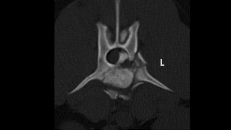
Figure 1: CT scan of the lumbar spine, showing a comminuted vertebral body and pedicle fracture of L3. Fracture fragments are visible within the spinal canal, compressing the spinal cord.
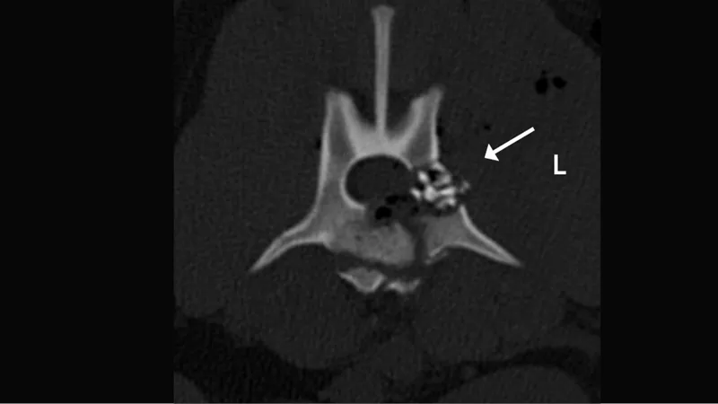
Figure 2: CT scan post surgery. The fragments compressing the spinal cord were removed and Xcyte is clearly visible within the fracture site (arrow).
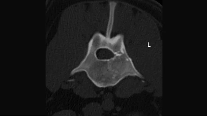
Figure 3: CT control 7 months after surgery showing a completely healed lumbar vertebra.
Vertebral fracture in a dog
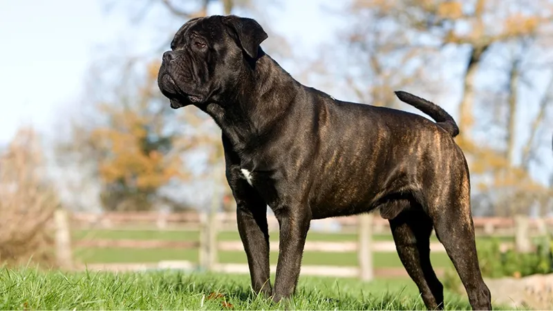
A one-year-old neutered male Cane Corso was brought to the small animal hospital after being hit by a car. He was lethargic and unable to walk. He presented with monoplegia with deep pain in the right hind limb and monopareses in the left hind limb. The tail was uniformly paralyzed. The neurological localization for the suspected vertebral damage was identified as the mid- to caudal-lumbar region. After initial stabilization, a CT scan was performed and revealed a highly comminuted compression fracture of the L3 vertebral body and pedicle on the left side. Multiple fracture fragments were visible within the spinal canal compressing the spinal cord (Figure 1).
Surgery was performed to remove the fracture fragments within the spinal canal. In addition, a new osteoinductive material, Xcyte, was placed at the fracture site to promote bone healing. First a small fat graft was inserted to protect the spinal cord from the new bone formation. Xcyte was then gently placed to fill the bone defect within the vertebral pedicle (Figure 2 ).
After surgery, the dog recovered well, was able to urinate on his own, and was discharged 5 days after initial presentation with improved tail motor. The left hind limb was still paralyzed with nociception that improved over the following weeks.
At a follow-up visit 7 months after surgery, the dog was able to walk normally and, according to the owner, was living a normal life with no neurological limitations. A control CT scan showed complete healing of the fracture site at this time (Figure 3)